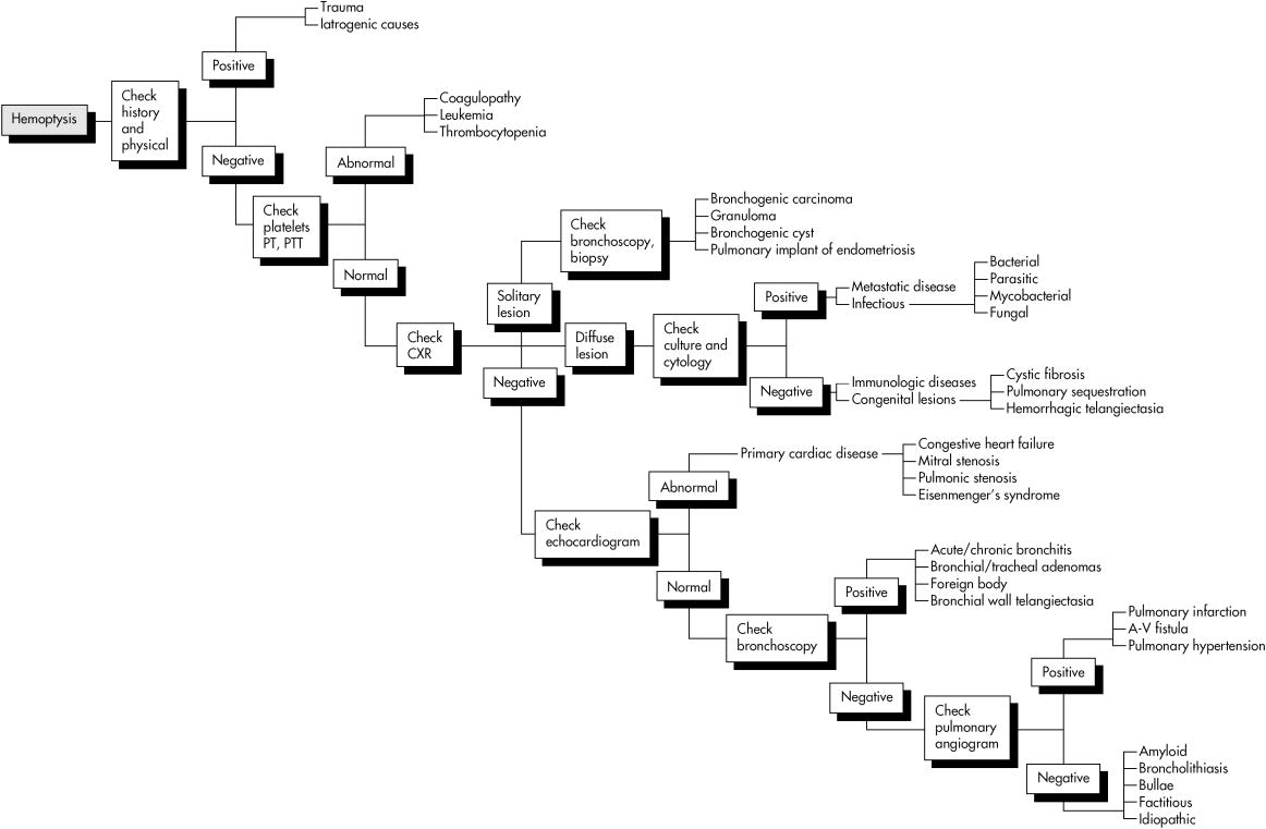
| Treatment of Hemoptysis:
* Localization of bleeding site by bronchoscopy or pulmonary
angiogram
1. Treat the identified underlying causes
!!!
Position good lung up
Cough suppression to prevent further coughing
which likely inhibits hemostasis.
Consider empirical antibiotics for lung infections
2. Bronchoscopic Management of Massive Hemoptysis
- Endotracheal Intubation
with endotracheal tube of large caliber for airway control & acute
respiratory distress
- to facilitate airway suctioning, ventilation, and bronchoscopy.
Selective intubation of the mainstem bronchi can be used to emergently secure
a blood-free airway in a patient with massive hemoptysis.
Double-lumen endotracheal tubes (DLTs) allow independent isolation of each
mainstem bronchus. However, there are problems with the use of DLTs
in patients with massive hemoptysis. Placement requires expertise, and the
DLT is difficult to secure in an unparalyzed patient.
-
Repeated bronchoscopic suctioning
-
Iced saline irrigation
-
Bronchoscopic Infusions or instillation of topical
Hemostatic or Vasoactive Drugs ( Materials)
Bronchoscopic application of epinephrine 1:1000,
1 to 2 mL, to the bleeding site sometimes slows the
bleeding.
Fibrinogen-thrombin mixtures have been used in Japan with good success. Although
commercial fibrinogen is not available in the United States for patient use,
cryoprecipitate-thrombin or thrombin alone remains a theoretically beneficial
alternative. Cold saline lavage through a rigid bronchoscope has also had
reported success, although controlled trials have not been done.
-
Bronchoscopic tamponade with Fogarty catheters
or Baloon tamponade.
- the balloon-tipped catheters to control massive hemoptysis.
Swan-Ganz catheters can also be used for this purpose, but their short length
requires they be carried on the exterior of the bronchoscope by a "bronchoscopic
shuttle" technique to facilitate bronchoscope removal. With the balloon inflated,
a less emergent decision regarding surgery can be made.
-
Tamponade with gauze or Gelfoam
-
Fibrin glue tamponade
-
Laser Photocoagulation (The Nd-YAG or argon
laser)
- is effective in controlling persistent hemoptysis from airway carcinomas
in approximately 60% of patients.
- Argon plasma coagulator
-
Electrocautery
-
A cryoprobe can be used via flexible
bronchoscope to freeze and coagulate the bleeding lesion.
-
Isolation of bronchial tree (double-lumen endotracheal tube)
-
Bronchoscopic brachytherapy
3. Selective Bronchial Artery Embolization
(BAE)
is useful in controlling most acutely bleeding patients
yet carries a high incidence of recurrent bleeding in some patient subsets.
The most difficult part of the procedure is to identify
the vessel(s) responsible for bleeding.
4. SURGERY: Thoractomy and resection
of the most involved segments or lobes of the lung.
With any of these techniques, coagulation of the bleeding lesion is more
important than resection of the lesion. If the bleeding is coming from distal
bronchi or pulmonary parenchyma, the distal tip of the flexible bronchoscope
is advanced as far as possible to tamponade the bronchus. This step alone
may keep the blood from flooding other bronchi and permit slow coagulation
of blood collected in the distal areas. Again, instillation of iced saline,
10 to 15 mL, through the bronchoscope may control the bleeding. Special
bronchial-blockade balloon catheters are available to tamponade distal bronchi.
The greatest danger from hemoptysis is from
asphyxiation rather than from exsanguination.
Bronchoscopic techniques are sometimes employed to control bleeding originating
in the tracheobronchial tree or pulmonary parenchyma.
Massive hemoptysis, defined as expectoration of
more than 200 mL of blood in a 24-hour period, is seen in less
than 5% of patients with hemoptysis. Treatment of significant hemoptysis
begins with diagnostic bronchoscopy to localize the site of bleeding; the
treatment approach is then selected.
Bronchoscopic techniques for management of massive hemoptysis are listed
as above .
Death occurs by asphyxiation and can occur very
suddenly.
Small volumes of blood or blood clots suddenly placed into the central airways
may necessitate endotracheal intubation even though the patient was speaking
in full sentences the minute before. Unfortunately, the frustrating inability
to know which patients will accelerate their hemoptysis suggests that all
patients with hemoptysis of greater than 200 mL be admitted to an area of
the hospital that can rapidly initiate airway support.
Major management issues are dependent on the rate
of bleeding.
Patients with 600 mL of hemoptysis in less than 4 hours carry up to a 71%
mortality rate vs. 22% and 5% if 600 mL of hemoptysis occurs in 4 to 16 hours
and 16 to 48 hours, respectively. Therefore precise quantitation of bleeding
requiring timed sputum containers at the beside is important. Therapy
for minor and moderate hemoptysis can usually proceed along diagnostic channels,
whereas massive hemoptysis must first focus on patient resuscitation and
stabilization .
When the Hemoptysis Stopped
-
Localization of bleeding site
-
Cough suppression to prevent further coughing which likely inhibits hemostasis.
-
Consider empirical antibiotics for lung infections
-
Diagnostic evaluation for underlying causes
REF: Mason: Murray & Nadel's Textbook
of Respiratory Medicine, 4th ed., 2005

 |