Acute Chemical Emergency
Stefanos N. Kales, M.D., M.P.H., and David C. Christiani, M.D., M.P.H.
NEJM
February 19, 2004, Volume 350:800-808
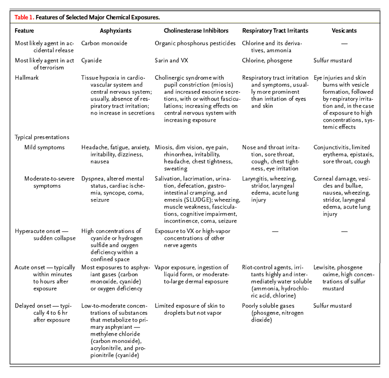
Acute chemical emergencies can occur as a result of an industrial disaster,1,2 occupational exposure,3,4 recreational mishap,5 natural catastrophe,6 chemical warfare,7,8 and acts of terrorism.9,10 This article reviews the health effects most commonly associated with the short-term release of industrial and environmental substances and with the use of chemical weapons. We emphasize the application of empirical principles and the recognition of four clinical syndromes, or "toxidromes," that are applicable to most scenarios of accidental release of chemicals and deliberate release as in acts of chemical terrorism. The classes of substances that correspond to these clinical syndromes are asphyxiants (e.g., cyanide), cholinesterase inhibitors (e.g., organophosphorus nerve agents), respiratory tract irritants (e.g., chlorine), and vesicants (e.g., mustard) (Table 1). The agents that cause each clinical syndrome require similar treatment.
Table 1. Features of Selected Major Chemical Exposures.

In accidental industrial releases, information about the presence of specific chemicals may be available from the personnel of the facility, safety officials, and other sources. In contrast, an act of terrorism is more likely to involve substances that cannot be immediately identified. Owing to the rapidity of the onset of similar symptoms in a group of persons or the close proximity of a group of persons to a release of hazardous materials, chemical exposures are more quickly recognizable than are exposures to biologic agents.11,12 However, in contrast to the period of latency that is associated with the effects of biologic agents, when serious chemical intoxication occurs, the window for effective therapy is often narrow. Furthermore, real-time identification of specific chemicals by means of environmental or clinical laboratory testing is difficult.13,14
General Principles
Empirical treatment of the casualties of an acute chemical emergency is of paramount importance. Treatment begins with ending the exposure, which can be accomplished by evacuating or extricating the affected persons and then by thorough decontamination. Persons in the vicinity of a chemical release that occurs outdoors can themselves take several simple steps. If they are outdoors, they should move away from the source of contamination — ideally, they should move upwind of it — until they reach an adequate shelter. If they are indoors, they should close all windows and doors and shut down both the heating and cooling systems, which could bring inside the contaminants that are outside. Persons who suspect that they have sustained an exposure to a chemical contaminant should remove and bag their clothing and shower thoroughly with soap and water as soon as possible.
Emergency personnel who approach the scene of the release of an unknown chemical should use portable radiation detectors in order to rule out the possibility that high levels of ionizing radiation are present.15,16 Affected persons within the zone contaminated by the release of a chemical can be extricated most safely by emergency personnel who are using the appropriate personal protective equipment. Early decontamination of persons who have been affected by the release of hazardous materials, before transport to a hospital, should be performed by trained first responders. Removing contaminated clothing can eliminate 85 to 90 percent of trapped chemical substances.11,13,17 After their clothing has been removed, injured persons should be irrigated with water, and then washed with soap and water; this approach is simplest and is effective in almost all situations.7,11,13,18
In a chemical emergency that involves a mass exposure, the majority of the affected persons are likely to be exposed minimally and to remain ambulatory and therefore able to reach a hospital by their own efforts. Thus, hospitals and triage centers should make advance preparation for shower facilities.13,19,20 In scenarios for the treatment of mass casualties, the projected number of persons affected by stress often exceeds the number of persons affected physically, by ratios ranging from 5:1 to 16:1.13,17,21,22 Therefore, resources to provide psychological support must be available both for casualties and for emergency personnel.
The clinical signs of severe chemical injury include altered mental status, respiratory insufficiency, cardiovascular instability, and a period of unconsciousness or convulsions. Initial supportive therapy should be focused on airway patency, ventilation, and circulation, at the same time that patients are examined for burns, trauma, and other injuries. When poisoning by industrial chemicals or chemical weapons is suspected, routine emergency guidelines should still be followed; these include considering the administration of naloxone to patients who have an altered mental status and respiratory depression.23 Some chemicals cause systemic intoxication, which may require treatment with antidotes. Early consultation with the regional poison center is recommended. Diazepam, cyanide antidote kits, atropine, and pralidoxime are the most important drugs to stockpile locally for the potential treatment of mass casualties of a chemical emergency.11,24
Asphyxiants
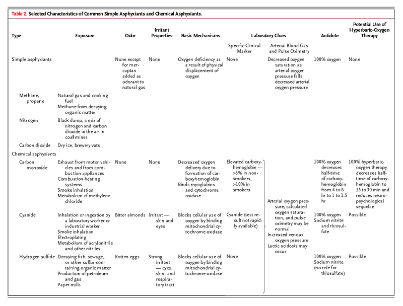
Asphyxiants are substances that cause tissue hypoxia with prominent neurologic and cardiovascular signs. Mild symptoms of asphyxia include headache, fatigue, dizziness, and nausea. More severe symptoms range from dyspnea, altered mental status, cardiac ischemia, and syncope to coma and seizure. Respiratory failure, if it occurs, generally results from depression of the central nervous system. Asphyxiants are classified as either simple or chemical on the basis of the mechanism of toxicity (Table 2). Simple asphyxiants (e.g., methane and nitrogen) physically displace oxygen in inspired air, and their inhalation results in oxygen deficiency and hypoxemia. Chemical asphyxiants (e.g., carbon monoxide, cyanide, and hydrogen sulfide) interfere with oxygen transport and cellular respiration and thereby cause tissue hypoxia. Therefore, in chemical asphyxiation, the partial pressure of arterial oxygen may not be reduced, but anaerobic metabolism often causes lactic acidosis.25,26,27,28
Table 2. Selected Characteristics of Common Simple Asphyxiants and Chemical Asphyxiants.

Carbon monoxide
![]()
![]()
is the most frequent cause of asphyxiant poisoning and the most common cause
of fatal occupational inhalation in the United States.29 The incidence of
carbon monoxide poisoning is greater during the winter season, because most
exposures result from the escape of the chemical from faulty heating systems
or in exhaust from combustion-powered vehicles or appliances and because
carbon monoxide readily accumulates indoors. A diagnosis of carbon monoxide
poisoning is confirmed by an elevated carboxyhemoglobin level, for which
there is a specific test with rapidly available results.25,30 Low
carboxyhemoglobin values must be interpreted cautiously, however, because
they can be the result of treatment with oxygen or substantial delays between
the end of the exposure and the carboxyhemoglobin measurement.25,31
Cyanide poisoning
![]()
![]()
should be suspected when a laboratory or industrial worker suddenly collapses.
Cyanides can have a secondary role in carbon monoxide poisoning that results
from smoke inhalation.32,33 The hallmarks of severe cyanide toxicity are
persistent hypotension and acidemia despite adequate arterial oxygenation.
Hydrogen sulfide poisoning produces a similar clinical picture. High
concentrations of hydrogen sulfide from decaying organic matter within a
confined space can rapidly "knock down" both initially exposed persons and
their would-be rescuers.4,34,35
For all cases of poisoning by asphyxiants, treatment begins with the administration of 100 percent oxygen. Oxygen reverses hypoxemia in cases of simple asphyxiation, accelerates the elimination of carbon monoxide,30,36 and helps to support persons poisoned by cyanide or hydrogen sulfide.26,34 In the United States, cyanide poisoning is treated with 100 percent oxygen along with sodium nitrite and thiosulfate, both of which are in the Lilly Antidote Kit.26,27,34,37 Nitrite induces the formation of methemoglobin, which is bound by cyanide, yielding cyanomethemoglobin. Because methemoglobin decreases the oxygen-carrying capacity of the blood, its levels must be monitored. Thiosulfate acts synergistically to accelerate the detoxification of cyanide to thiocyanate. Adverse effects are rare, and thiosulfate can be given safely when cyanide poisoning is suspected.34 For cyanide poisoning due to smoke inhalation, most authorities recommend the use of thiosulfate, oxygen, and supportive measures and recommend reserving nitrites for patients who are hypotensive, acidemic, or comatose.32,37,38 Nitrite-induced methemoglobinemia aggravates the decrease in oxygen-carrying capacity that is due to carboxyhemoglobinemia.
For the treatment of poisoning by hydrogen sulfide, which preferentially binds methemoglobin, the administration of 100 percent oxygen and sodium nitrite is recommended.34,39,40 Thiosulfate, however, is not indicated in cases of hydrogen sulfide poisoning. Hydrogen sulfide is highly irritating, and patients must be monitored for ophthalmic toxicity ("gas eye") and acute lung injury.40,41
Additional treatment with hyperbaric oxygen may be offered to selected patients with chemical asphyxia. Hyperbaric oxygen accelerates the elimination of carbon monoxide and decreases the frequency of the cognitive sequelae that can result from severe exposure to carbon monoxide.36,42 It may also be beneficial in the treatment of poisoning by cyanide and hydrogen sulfide.43,44,45 The role of hyperbaric-oxygen treatment in a mass exposure to asphyxiant substances is restricted by the limited availability of hyperbaric chambers.
Organic phosphorus pesticides, carbamate pesticides, and the organophosphorus compounds that are developed as weapons known as "nerve agents" (e.g., sarin, soman, tabun, and VX) all inhibit acetylcholinesterase, resulting in cholinergic overstimulation, with both muscarinic and nicotinic effects.46,47,48 Muscarinic symptoms include profuse exocrine secretions (tearing, rhinorrhea, salivation, bronchorrhea, and sweating), in addition to ophthalmic symptoms, such as miosis, dim vision, headache, and eye pain. Exposure to large doses of cholinesterase inhibitors, especially if these are ingested, may cause abdominal cramping, nausea, emesis, diarrhea, and fecal and urinary incontinence. Nicotinic symptoms include weakness of the skeletal muscles, fasciculations, and paralysis. Cardiovascular effects of poisoning are mixed, but initially tachycardia and hypertension due to nicotinic stimulation usually predominate.48,49 Effects on the central nervous system range from irritability and mild cognitive impairment to convulsions and coma.48,50,51 Multiple mechanisms (e.g., hypersecretion, bronchoconstriction, thoracic weakness, and decreased respiratory drive) can contribute to respiratory failure. Depression of erythrocyte cholinesterase and serum cholinesterase activity provides confirmation of severe intoxication.9,48,51,52 However, treatment cannot await the results of testing of cholinesterase activity because tests results are not rapidly available.11,13
Cholinesterase inhibitors are absorbed by inhalation, by ingestion, and through the skin, and they may contaminate emergency personnel who are inadequately protected.47,50 Supplemental oxygen, suctioning, and mechanical ventilation may be needed to support the patient. Three antidotes — atropine, pralidoxime, and diazepam — are useful. Atropine works primarily at muscarinic sites. It is administered to adults in doses of 2 mg every 5 to 10 minutes, and the dose is adjusted to minimize dyspnea, airway resistance, and respiratory secretions.48,52 Pralidoxime reactivates acetylcholinesterase and works at nicotinic, muscarinic, and central nervous system receptors. The initial dose of pralidoxime is 1 g administered intravenously over a period of 20 to 30 minutes.7,52
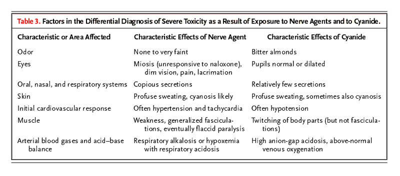
Benzodiazepines are the only effective anticonvulsant drugs for the treatment of persons poisoned with nerve agents and should be administered to all persons with severe intoxication by such agents (i.e., patients with seizure, loss of consciousness, or toxic effects in two or more organ systems).7,17,48 In an instance of terrorism in which persons suddenly collapse with coma, seizure, or apnea, cyanide is the other chemical agent to be considered.11,17,53 Persons affected by nerve agents or cyanide require airway support and the administration of 100 percent oxygen. Seizures in both cases should be treated with benzodiazepines. Characteristics that differentiate the diagnosis of poisoning by cyanide from that of poisoning by nerve agents are listed in Table 3.
Table 3. Factors in the Differential Diagnosis of Severe Toxicity as a Result of Exposure to Nerve Agents and to Cyanide.

There are several differences between organophosphorus nerve agents and structurally related organic phosphorus insecticides. The insecticides are oily, less volatile liquids. They have a slower onset of toxicity, but their effects last longer and require a larger cumulative dose of atropine.46,50,53 Nerve agents are watery and volatile, acting rapidly and severely, but their effects last for a shorter time and require a smaller total dose of atropine.7,53 Over time, organophosphorus–acetylcholinesterase binding becomes irreversibly covalent and resistant to reactivation by pralidoxime, in a process known as "aging." Aging has clinical implications for soman, which ages in minutes, and sarin, which ages over a period of three to five hours.50,54 Pralidoxime should never be withheld, however, out of concern that it might be administered too late after exposure.54 For organophosphorus insecticides, aging is not clinically relevant because these agents age at a slow rate.47 Among nerve agents, VX has several unique characteristics. It is oily, is persistent in the environment, and ages minimally, but even one drop of the substance on the skin can be lethal.52
Carbamate insecticides
have a more limited penetration of the central nervous system, inhibit
acetylcholinesterase reversibly, and result in a shorter, milder course than
organophosphorus compounds. Nevertheless, in the treatment of severe cholinergic
syndromes, it is prudent to use both atropine and pralidoxime.47
The hazardous materials most frequently released in industrial accidents are irritants to the respiratory tract.55,56,57 Other respiratory tract irritants are tear gas and choking agents that are used in warfare. Direct tissue reactivity, reflex stimulation, water solubility, and dose are factors that determine the clinical effects of these substances. Highly soluble irritants, such as ammonia, are absorbed in the upper respiratory tract, where symptoms develop that are early warnings of toxicity, whereas less soluble irritants, such as phosgene, penetrate more deeply and may cause acute lung injury with a delayed onset.7,58 Regardless of the degree of solubility of the chemical irritant, however, any massive exposure may have severe effects on the upper respiratory tract (e.g., laryngeal edema) or the lower respiratory tract (e.g., acute lung injury).7,58,59
The chemical agents used for riot control — tear gas or other "lacrimators" — are aerosolized solids that cause intense, immediate, and usually self-limited burning on exposed body surfaces, especially the eyes.60 Ammonia, hydrochloric acid, sulfuric acid, and the chloramines — which, in a common mistake, are produced by the inappropriate mixing of ammonia and household bleach (hypochlorite) — are highly soluble irritants to the upper respiratory tract.58,61 In the case of tear gas and the highly soluble agents, intense or prolonged exposure or the presence of underlying lung disease may result in bronchospasm and even acute lung injury.58,62 Chlorine has an intermediate solubility; in small doses it irritates the upper respiratory tract, whereas in larger doses it leads to bronchospasm and eventually to acute lung injury.63
Phosgene is the prototypical low-solubility irritant.7 It irritates the mucosa, but neither the irritation nor the odor of the substance (which has been likened to new-mown hay, moldy hay, or green corn) provides an adequate warning of its presence.59,64 As late as 15 to 48 hours after the exposure, acute lung injury may be manifested in persons who were previously asymptomatic.59,64 Dyspnea or radiographic evidence of pulmonary edema within four hours after exposure to phosgene indicates a worse prognosis and requires treatment in an intensive care unit.7,59 In persons who remain asymptomatic and whose lungs appear clear on chest films obtained eight hours after exposure, acute lung injury is unlikely to develop.64 Nitrogen dioxide is another poorly soluble gas. Silo-filler's disease can develop in agricultural workers who inhale high concentrations of the nitrogen dioxide that may accumulate inside silos.65
Treatment of the effects of respiratory irritants begins with life support, the administration of high-flow oxygen, and decontamination by irrigation of the eyes and skin. An assessment of the severity of the effects is based on the particular substance or substances involved, the duration of the exposure, and a determination of whether the patient was exposed to the substance within a confined space and whether there was loss of consciousness. Patients in whom hoarseness, stridor, upper-airway burns, wheezing, or altered mental status develop may require endotracheal intubation. Bronchodilators are indicated to treat bronchospasm, and corticosteroids may be added as therapy for severe airway reactivity.7,60 Nebulized bicarbonate has been advocated as therapy to neutralize chlorine derivatives, but data from controlled studies of its efficacy are lacking.66
For acute lung injury, the treatment remains supportive. Between the exposure and the onset of symptoms, bed rest is crucial, because physical exertion exacerbates inflammation of the lungs.7,17,59,64 The use of corticosteroids has been recommended for possible prophylaxis and as therapy.59,67 Although their use in the treatment of the effects of exposure to phosgene is controversial,7,17 corticosteroids are favored for the treatment of moderate-to-severe exposure to nitrogen dioxide.17,68 Positive end-expiratory pressure may be used to help maintain oxygenation in the presence of pulmonary edema.59,64 The administration of diuretics should be avoided because they may aggravate intravascular hypovolemia.7,64 Although bacterial superinfection is a recognized complication of acute lung injury due to phosgene, the prophylactic use of antibiotic drugs is not recommended.59
Vesicants, which are blistering agents, are extremely irritating to the eyes, skin, and airways.69,70,71 Mustard is the most important agent in this class, and its use historically has caused the greatest number of casualties of all chemical warfare agents.72 Mustard is a liquid at room temperature, but it becomes a vapor hazard as the ambient temperature rises.69,73 Affected persons and clinicians may be misled by the typical period of latency of 4 to 12 hours between exposure and the onset of symptoms and may therefore not initially recognize that an exposure occurred.17,74 Within minutes, however, absorbed mustard becomes fixed in the dermis or penetrates the circulation.7,75 Therefore, decontamination, which is always indicated in persons who have been exposed to mustard, is most effective when performed immediately after exposure.53,69
Mustard is a radiomimetic alkylating agent that affects DNA chains69,72 and is an inflammatory activator.7,17 The cutaneous and ophthalmic effects of exposure to mustard are the most prominent and the first to appear. Ophthalmic effects range from conjunctivitis to corneal damage and can include temporary or permanent loss of vision.69,76 Dermatologic lesions can develop and progress from erythema to vesicles and bullae with a predilection for forming in intertriginous areas.73,74,75 Airway involvement can occur, usually within 24 hours after the exposure and can range from epistaxis, pharyngitis, laryngitis, and cough to dyspnea and sputum production to hemorrhagic edema, the formation of a pseudomembrane, and mucosal sloughing with possible airway obstruction.7,72,74 Pulmonary complications are the most common cause of death from exposure to mustard.74,75 High doses affect rapidly dividing cells69 and usually result in nausea and vomiting.74 Within days to weeks after exposure to mustard, hematopoietic suppression may be manifested as leukopenia,74,76,77 although it can be initially obscured by a reactive leukocytosis.69 An exposure that may be fatal is indicated by effects on the patient's airway within six hours,7,75 burns over more than 25 percent of the total body surface,17,74 and an absolute white-cell count of less than 200 per cubic millimeter.69,75
After immediate decontamination and eye irrigation, persons affected by a severe exposure to mustard require supportive measures, including pulmonary care similar to that used to treat patients affected by respiratory tract irritants. In addition, such persons may require specialized ophthalmic treatment,76 burn care,77 and critical care.69 Early ophthalmic treatment consists of the administration of topical anticholinergic agents, antibiotics, and petrolatum to prevent the eyelids from sticking.75,76 Care of burns involves débridement, the application of topical antibiotic medication, and liberal administration of analgesic drugs.7,77 Large blisters should be unroofed with care.74 The blister fluid does not contain active mustard.77 Overhydration should be avoided in patients with mustard burns, who have less fluid loss than patients with thermal burns.11,67
Although there are no antidotes to mustard, emerging evidence suggests that early treatment with nonsteroidal antiinflammatory drugs may be beneficial.17,78,79 The use of thiosulfate has been shown to decrease systemic toxicity and mortality in animals.7,69 In the presence of hematopoietic suppression, the use of nonabsorbable antibiotics to sterilize the gut may prevent enteric sepsis.7 Granulocyte colony-stimulating factor should be considered for the treatment of severe neutropenia.11,67
Most chemical burns of the skin are due to contact with simple acids or bases. Unlike mustard, these substances are not considered vesicants, and most burns due to them do not result in systemic toxicity. The primary treatment is decontamination, including vigorous irrigation with water. Burns from hydrogen fluoride, which is a component of some household rust removers and is used in certain industries, require special treatment. Hydrofluoric acid releases fluoride, which has a high affinity for calcium and magnesium. Dilute exposures result in delayed symptoms of severe pain, whereas extensive burns and exposure to high concentrations can result in life-threatening hypocalcemia and hypomagnesemia80,81 and may require the administration of local and parenteral calcium preparations.80
Community Preparedness
In all serious cases of exposure to chemical substances, a successful outcome hinges on the extrication of casualties, immediate provision of basic life support and decontamination, and follow-up with excellent supportive care. Community preparedness for possible toxic chemical releases requires well-organized emergency-medical-response systems, as well as clinicians and hospitals trained for readiness. Emergency planning should be applicable to both accidental and deliberate chemical releases.
Supported by a grant (ES00002) from the Environmental Health Center, National Institutes of Health, and by the Department of Environmental Health, Harvard School of Public Health.
We are indebted to Dr. Thomas Glick for his insightful comments.
From the Cambridge Health Alliance, Harvard Medical School, Cambridge, Mass. (S.N.K.); the Department of Environmental Health, Occupational Health Program, Harvard School of Public Health, Boston (S.N.K., D.C.C.); the Pulmonary–Critical Care Unit, Massachusetts General Hospital and Harvard Medical School, Boston (D.C.C.); and the Northeast Specialty and Rehabilitation Hospital–Center for Occupational and Environmental Medicine, Braintree, Mass. (D.C.C.).
Address reprint requests to Dr. Kales at the Cambridge Health Alliance, Department of Medicine, Occupational and Environmental Health, 1493 Cambridge St., Cambridge, MA 02139, or at skales@challiance.org.
References