Status Asthmaticus in Adults
See
Asthma
REF: UpToDate 2006 |
| Assessment |
Management |
Hospitalization | Useful
algorithms | Recommendation
|
| INTRODUCTION
— Severe attacks of asthma poorly responsive to adrenergic
agents and associated with signs or symptoms of potential respiratory failure
are often referred to as "status
asthmaticus." The term is now applied to asthma attacks of
unusual severity.
|
|
| PATHOGENESIS
The pathologic changes found on post-mortem examination of airways
from most patients dying of severe asthma differ only in severity from those
found in patients with mild asthma who die of other causes:
-
Bronchial wall thickening from edema and inflammatory cell infiltration.
-
Hypertrophy and hyperplasia of bronchial smooth muscle and submucosal glands.
-
Deposition of collagen beneath the epithelial basement membrane.
-
In addition, most asthma deaths are associated with prominent intraluminal
inspissation of secretions, leading to occlusion of up to 50 percent of the
total cross sectional area of airways two mm in diameter.
-
In some deaths, however, bronchial mucus is absent, suggesting that airway
obstruction was predominantly due to intense smooth muscle contraction.
These pathologic patterns may correlate with differences in the rate of
progression of attacks:
-
A rapid response to bronchodilator therapy of "acute asphyxic asthma," in
which the interval from the appearance of symptoms to intubation is less
than three hours, has been interpreted as suggesting that bronchial smooth
muscle contraction is the predominant cause of airway narrowing.
-
The slower response to therapy of asthmatic exacerbations that progressed
over many hours, days, or even weeks, may indicate greater contributions
from mucus inspissation and inflammatory thickening of the bronchial wall.

|
| Assessment |
Management |
Hospitalization | Useful
algorithms | Recommendation
|
| ASSESSMENT
OF SEVERITY
History
— The most powerful predictive feature in the history that
a severe episode may be life-threatening is any history of prior intubation
and mechanical ventilation for asthma. In comparison, progression of symptoms
despite treatment with high doses of inhaled beta-agonists and oral or inhaled
corticosteroids probably indicates a greater risk of a poor or slow response
to initial emergency department therapy; it does not, however, alter
recommendations for the initial therapy given.
Physical examination
— Gross physical findings of severe asthma include any alteration
in consciousness, fatigue, upright posture, diaphoresis, and the use of accessory
muscles of breathing. Tachypnea (>30/min), tachycardia (>120/min),
and an inspiratory fall in systolic blood pressure (pulsus paradoxus) of
more 15 mmHg are also more common in severe attacks. It is important
to appreciate, however, that the absence of these findings does not exclude
even immediately life-threatening airflow obstruction, especially if the
patient is exhausted or obtunded.
Examination of the chest typically reveals overinflation, reduced respiratory
excursion, and diffuse expiratory wheezing, but the pitch or intensity of
wheezing does not discriminate between degrees of severity. Careful
inspection of the mouth and pharynx and auscultation over the upper airway
may provide clues that the site of obstruction is in the upper airway, as
from epiglottitis, angioedema, or vocal cord dysfunction.
Measurement of peak expiratory flow rate
— The most direct assessment of airflow obstruction is measurement of
spirometry or peak expiratory flow rate (PEFR). However, some patients
are too dyspneic to perform even this test until bronchodilator therapy has
been given. Whenever possible, the PEFR should be measured initially to provide
a baseline and at successive intervals during treatment. Predicted values
differ with size and age, but a peak flow below 120 L/min or an FEV1 below
1.0 liters indicates severe obstruction for all but unusually small adults.
Arterial blood gas measurements
— The most direct assessment of the impact of airflow obstruction on
ventilation is measurement of arterial blood gases. The critical information
lies not in arterial oxygen tension, for severe hypoxemia is rare
in asthma and the customarily modest hypoxemia is responsive to modest oxygen
supplementation. Far more important is the arterial PCO2.
Respiratory drive is almost invariably increased in acute asthma, resulting
in hyperventilation and a correspondingly decreased PaCO2. Thus,
an elevated or even normal PaCO2 indicates that
airway narrowing is so severe that the ventilatory demands of the respiratory
center cannot be met. Respiratory failure can then develop rapidly
with any further bronchoconstriction or respiratory muscle fatigue.
Because CO2 retention develops
only in severe asthma and oxygen saturation can be measured by pulse oximetry,
arterial blood gases need not be measured in most asthmatic patients presenting
for emergency care. Furthermore, even patients with CO2 retention usually
respond to aggressive drug therapy, so it could be argued that
arterial blood gases need only be determined on initial assessment in
the following cases:
-
Patients gasping for air.
-
Patients unable to speak more than two or three words.
-
Patients with obtunded consciousness or in cardiopulmonary arrest.
-
Patients with severe asthma who respond poorly to initial aggressive therapy.
-
Repeated evaluation after initial treatment may be even more important than
the initial assessment of severity. The response to the first two hours of
treatment appears to be the most powerful predictor of outcome.

|
| Assessment |
Management |
Hospitalization | Useful
algorithms | Recommendation
|
| Management of
Status Asthmaticus
* Correction of any
precipitating causes!
-
Oxygen supplement
-
Bronchodilators
a. Inhaled Beta agonists as
albuterol (Ventolin/Proventil)
- It is most commonly given by handheld nebulizer, at a dose of 2.5
mg dissolved in two mL of isotonic saline.
- In patients with particularly severe obstruction, nebulized albuterol may
be given continuously until the obstruction is improved or toxicity (tachycardia,
arrhythmias, skeletal muscle tremor) supervenes.
- The modest fall in the plasma potassium concentration of
about 0.7 meq/L caused by intensive beta-agonist therapy may induce slight
Q-T prolongation on the electrocardiogram. However, this translocation of
potassium into the cells is infrequently of practical importance except for
patients who are hypokalemic from other causes or who are taking digitalis.
Similar modest falls in the plasma magnesium and phosphate concentration
also occur via the same mechanism.
b. Ipratropium bromide (Atrovent™)
by inhalation
- As a practical matter, it appears that the addition of 0.25 mg of
ipratropium to a large dose of albuterol (five mg) in the solution given
by nebulizer results in greater improvement in FEV1 than does albuterol
alone (26 versus 20 percent in one study)
- For patients requiring frequent bronchodilator aerosol treatment for several
hours, alternating albuterol and ipratropium treatments offers a way of
maintaining bronchodilation while reducing the risk of beta agonist
toxicity.
c. Theophylline by intravenous infusion
adds to the bronchodilation achieved by maximal doses of an inhaled beta
agonist.
- Most short-term studies show that intravenous infusion of theophylline
(aminophylline) adds toxicity but no further immediate bronchodilation to
that achieved by nebulized therapy with a beta agonist. Furthermore, most
longer-term studies show that addition of intravenous theophylline to inhaled
beta agonist and intravenous corticosteroid therapy has no influence on the
course or duration of hospitalization
-
Corticosteroids - dramatically reduces
the need for hospitalization and should be instituted in patients who do
not respond immediately to bronchodilators.
- reducing mucosal edema and inflammatory cell infiltration; and decreasing
mucus secretion
- for example, IV methylprednisolone (125
mg) given on presentation to the emergency department was associated
with a hospitalization rate of 19 percent compared to 47 percent in the placebo
treated group.
- the current practice of methylprednisolone 2 mg/kg
IV q6hr. However, a meta-analysis of 30 randomized trials suggests
that half that dose is probably maximally effective.
- PO 60 mg of prednisone q6hr appears to as effective as intravenous
methylprednisolone.
Once oral corticosteroids are started, they should be continued for
at least one week, and the patient should be seen in follow-up before the
dose is tapered or discontinued. It was once common practice to reduce
the dose gradually, such as a decrease of five mg of prednisone every other
day over seven to ten days. However, there appears to be little risk in abruptly
stopping short courses (ie, less than 10 days) of oral therapy in patients
whose symptoms, physical findings, and peak flow have normalized.
- both 40 mg and 125 mg of methylprednisolone IV q6hr caused a
significantly more rapid improvement in FEV1 than did 15 mg.
- Inhaled corticosteroids may take several days for their effects to become
apparent, and possibly several months before they reach a plateau [49]. As
a result, inhaled therapy should be started before oral therapy is discontinued.
-
Parenteral magnesium
— The two largest prospective, double blind, controlled studies reported
thus far showed that magnesium sulfate added little or no further bronchodilation
to that achieved with an inhaled beta agonist. However, the numbers of subjects
studied were too few to definitely exclude possibly responsive subgroups,
such as premenopausal women.
_ The dose of magnesium typically given, 2 gm IV over 20
minutes, has little toxicity and is unlikely to harm a patient with
severe asthma. Nevertheless, we cannot recommend magnesium sulfate until
there is evidence that at least some patients benefit from its addition to
usual therapy.
-
Antibiotics — Viral but not bacterial
respiratory infections are common precipitants of asthma exacerbations. It
is therefore not surprising that amoxicillin has been shown not to affect
the course of acute asthmatic attacks. Antibiotics are now reserved for patients
with fever, leukocytosis (>15,000/mm3), and a pulmonary infiltrate on
the chest radiograph.
-
Intubation & Ventilator
- The decision to proceed to intubation and mechanical ventilation
should be made before the patient is in extremis. Asthmatics have exaggerated
bronchial responsiveness, and intubation is difficult in any circumstance,
let alone under the conditions of hypoxia, hypercapnia, and acidosis of
respiratory failure. There are no explicit and inclusive guidelines as to
when intubation should be performed; the decision to proceed is best based
on an integrative clinical assessment of the patient's ability to continue
ventilation until therapy becomes effective.
Worsening fatigue and persistent or increased
hypercapnia weigh heavily in favor of intubation.
The initial goal of therapy is simply to reverse obstruction to airflow,
as documented by improvement in the peak flow or FEV1 obtained 1 to 2 hours
after therapy is started. The ultimate goal is a sustained improvement in
either measurement to >70 percent of predicted or the patient's personal
best. This should be accompanied by a decrease in symptoms, so that the patient
is no longer short of breath at rest or with minimal exertion.

|
| Assessment |
Management |
Hospitalization | Useful
algorithms | Recommendation
|
REASSESSMENT
AND HOSPITALIZATION
— The severity of the asthmatic attack should be continually assessed
in the Emergency Department on at least an hourly basis after initiating
therapy. Most acute exacerbations of asthma respond well to therapy with
an inhaled beta agonist and a systemic corticosteroid. Prompt resolution
of symptoms and improvement in peak flow to greater than 70 percent of the
predicted value permits the patient to be sent home. However, bronchospasm
may recur, even in patients with such prompt and nearly complete improvement.
This is most likely to occur in the next 72 hours. As a result, the patient
should be given clear instructions for an "action plan" to treat recurrent
symptoms or a fall in peak flow.
The course is different for the many patients who do not respond so completely.
In this setting, the decision about the need for hospitalization is usually
made after four to six hours of therapy. Patients with an incomplete or poor
response to therapy at this time should be considered for admission.
Clear indications for admission are:
-
Fatigue.
-
Persistence of severe dyspnea.
-
Peak flow persistently below 50 percent predicted.
-
Pneumothorax or pneumomediastinum.
-
History of previous intubation for asthma.
-
Comorbidity from coronary artery disease.
For patients with better, but still incomplete responses, the decision about
hospitalization must weigh other considerations, such as the history of prior
attacks, the home situation, and the patient's skills in self-assessment
and treatment.
Indications for admission to an intensive or
intermediate care unit are persisting signs of severity:
-
Use of accessory muscles.
-
Fatigue
-
Peak flow under 150 L/min.
-
Normal or elevated arterial PCO2.
-
Marked increase in dyspnea or a fall in peak flow after a maximal expiratory
maneuver, eg, when performing the peak flow test.

|
| Assessment |
Management |
Hospitalization | Useful
algorithms | Recommendation
|
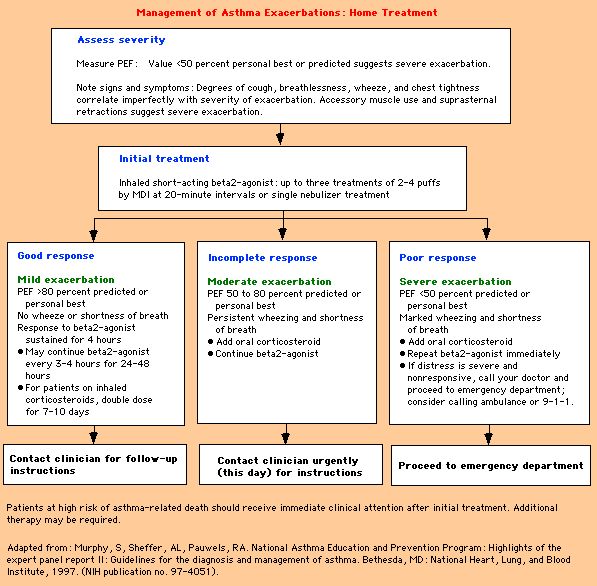
| Home Management
of Asthma Exacerbation


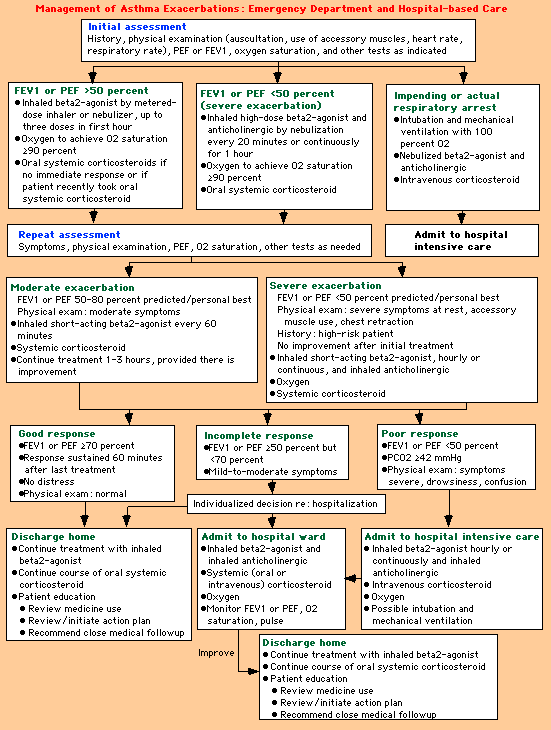
Hospital Management of Asthma
Exacerbation


|
| Assessment |
Management |
Hospitalization | Useful
algorithms | Recommendation
|
RECOMMENDATIONS
— Useful algorithms for managing acute exacerbations of asthma in the
home and in the Emergency Department setting have been published by the National
Asthma Expert Panel (show figure 5 and show figure 6). The basic principles
of care are to:
-
Carefully assess the severity of the attack.
-
Use inhaled beta agonists early and frequently.
-
Start oral or intravenous corticosteroids early if there is not an immediate
response to beta agonists.
-
Make frequent (every one to two hours) objective assessments of the response
to therapy until clear, sustained, improvement is documented.
-
Admit patients who do not respond well after four to six hours to a setting
of high surveillance and care.
-
Educate patients about the principles of self-management for early recognition
and treatment of a recurrent attack (develop an "action plan" for recurrent
symptoms).
|
Medications for Asthma & COPD

 |
Beta-2 Agonist
* * Aerochamber - better delivery. Spacer device
Inhaled short-acting beta2
agonists:
first-line p.r.n. therapy;
no significant differences in efficacy among inhaled short-acting
beta2 agonists; dilute aerosols to minimum of 4 ml at gas
flow of 6-8 L/min
-
Albuterol (Ventolin, Proventil)
-
Nebulizer, 5 mg/ml 0.5 ml in 2.5 ml NS(2.5
mg)
-
Dose: maintenance, 1.25–5.0 mg
q. 4–8 hr p.r.n; exacerbation, 5 mg q. 2 hr
-
MDI, 84 ¼g/puff:
MDI as effective as nebulizer when
used with spacer
-
Dose:
maintenance,
2–4 puffs q. 6 hr p.r.n; exacerbation, 3–8 puffs q. 2
hr
-
Pirbuterol (Maxair)
-
MDI, 200 ¼g/puff
-
Dose: maintenance, 2–4 puffs q. 6 hr p.r.n.;
exacerbation, not studied
-
Alupent 0.3 ml in 2.5 ml
NS (15 mg) (Metaproterenol)
Combined short-acting beta2 agonist
and anticholinergic:
use when both are indicated
-
MDI
Combivent albuterol (90 ¼g/puff)
+ ipratropium bromide (18 ¼g/puff)
Dose: maintenance, not recommended;
exacerbation, 3–8 puffs q. 2 hr
Inhaled long-acting beta2 agonists:
first-line scheduled bronchodilator
therapy
-
Salmeterol
(Serevent Diskus): Slower onset of action;
not used as a rescue bronchodilator
-
MDI, 21 ¼g/puff
-
Dose: maintenance, 2 puffs q. 12 hr;
exacerbation, not recommended
-
Cost/mo: $50.00–69.99
-
DPI, 50 ¼g/blister
-
Dose: maintenance, 1 blister q. 12 hr
-
Formoterol
(Foradil)
: faster onset of action than
salmeterol; may be used as a rescue bronchodilator
-
DPI, 12 ¼g/capsule
-
Dose: maintenance, 1 capsule q. 12 hr; exacerbation,
not recommended
Combined long-acting beta2 agonit and
corticosteroid:
-
Advair (fluticasone/Flovent +
salmeterol/Serevent) DPI 100/50, 250/50, 500/50) 1 puff bid
 |
Inhaled
Corticosteroids:
first choice of anti-inflammatory agents; various
inhaled corticosteroid agents differ in potency
-
Fluticasone (Flovent): highest
potency; also formulated in combination with salmeterol in DPI
-
Dose: low, 83-264 µg; medium, 264-660 µg;
high, > 660 µg
-
44 µg/puff: low, 2-4 puffs/day
-
110 µg/puff: low, 2 puffs/day; medium, 2-6
puffs/day; high, > 6 puffs/day
-
220 µg/puff: medium, 1-2 puffs/day; high,
> 3 puffs/day
-
Budesonide (Pulmocort): second-highest
potency; DPI inhaler
-
Dose: low, 200-400 µg; medium, 400-600 µg;
high, > 600 µg
-
220 µg/puff: low, 1-2 puffs/day; medium,
2-3 puffs/day; high, > 3 puffs/day
-
Beclomethasone (QVAR): third-highest
potency; available in HFA MDI, possibly improving effectiveness
-
Dose: low, 168-504 µg; medium, 504-840 µg;
high, > 840 µg
-
42 µg/puff: low, 4-12 puffs/day; medium,
12-20 puffs/day; high, > 20 puffs/day
-
40-80 mcg 1-2 puffs bid
-
Flunisolide (Aerobid): lowest potency
-
Dose: low, 500-1,000 µg; medium, 1,000-2,000
µg; high, > 2000 µg
-
42 µg/puff: low, 4-12 puffs/day; medium,
12-20 puffs/day; high, > 20 puffs/day
-
Triamcinolone (Azmacort): lowest potency;
provided with spacer
Dose: low, 400-1,000 µg; medium, 1,000-2,000
µg; high, > 2,000 µg
100 µg/puff: low, 4-10 puffs/day; medium,
10-20 puffs/day; high, > 20 puffs/day
Systemic Corticosteroids
-
Prednisone: usual oral agent; q.d. initially, then wean off, if possible,
or switch to q.o.d.
-
Dose: initial, 0.5-1.0 mg/kg/day; maintenance, none or minimal
-
Methylprednisolone, oral: less
commonly used oral agent; q.d. initially, then wean off, if possible,
or switch to q.o.d.
-
Dose: initial, 24-48 mg/day; maintenance, none or minimal
-
Methylprednisolone, I.V.:
usual I.V. agent; oral therapy is as effective
-
Dose: initial, 60-125 mg IV q. 6-8 hr;
maintenance, none
 |
| Anticholinergic
drugs:
Atrovent (Ipratropium) inhaler 2-4 puffs
q6h
Atrovent 0.5mg (1 unit dose) nebulizer soln in 2.5 mL NS
3-4x/d
Atropin 0.5 mg in 2.5 ml NS q6-8h
 |
Leukotriene
Inhibitors:
less effective than inhaled corticosteroids; help with associated allergic
rhinitis; use for aspirin-sensitive patients
-
Montelukast (Singulair): first choice
of leukotriene inhibitors; note q.h.s. dosing; no lab monitoring or restrictions
related to meals
-
Dose: 10 mg q.h.s.
-
Cost/mo: $60.00-69.99
-
Zafirlukast (Accolate): should
be taken at least 1 hr before or 2 hr after meals
-
Dose: 20 mg b.i.d.
-
Cost/mo: $50.00-59.00
-
Zileuton (Zyflo): must monitor
LFTs
-
Dose: 600 mg q.i.d.
-
Cost/mo: $79.00-89.99
-
Methotrexate (Rheumatrex, Trxall): efficacy
controversial; toxic drug, must monitor blood counts and LFTs closely; should
be given only by asthma expert
-
Dose: 7.5 mg/wk (adjust to effect; maximum 25
mg/wk)
-
Cost/mo:
$10.00-19.99
 |
| OTHERS RX:
Cromolyn Sodium
(Intal): much less potent than
inhaled steroids; used more often in children; no steroid side
effects
-
Dose: initial, 2-4 puffs q.i.d.; maintenance,
2 puffs q.i.d.
-
Cost/mo:
$30.00-39.99/canister
Nedocromil:
much less potent than inhaled steroids; no steroid side
effects
-
Dose: initial,
2 puffs
q.i.d.; maintenance,
2 puffs q.i.d.
Nonselective beta-2 agonists
-
* Patients whose bronchoconstriction is resistant to continuous handheld
nebulizer treatments with traditional beta-2 agonists may be candidates for
nonselective beta-2 agonists (eg, epinephrine 0.3-0.5
mg SC q 30 min x 3; or
terbutaline 0.25 mg SC) administered
subcutaneously. However, systemic therapy has no proven advantage over aerosol
therapy with selective beta-2 agents.
Susphrine 0.1-0.3 ml 1:200 SQ
*Avoid or use cautiously in heart or HBP pts.
* Aminophylline
The loading dose is usually 5-6 mg/kg, followed
by a continuous infusion of 0.5-0.9 mg/kg/h
-
o Conflicting reports on the efficacy of aminophylline therapy have made
it controversial.
-
o Starting intravenous aminophylline may be reasonable in patients who do
not respond to medical treatment with bronchodilators, oxygen, corticosteroids,
and intravenous fluids within 24 hours.
-
o Recent data suggest that aminophylline may have an anti-inflammatory effect
in addition to its bronchodilator properties.
-
o The loading dose is usually 5-6 mg/kg, followed by a continuous infusion
of 0.5-0.9 mg/kg/h.
-
o Physicians must monitor a patient's theophylline level. Traditionally,
the level was targeted to the higher end of the local therapeutic range;
however, many authorities suggest that the lower portion of the range (ie,
>5 but <10) may be preferable if the patient can obtain the benefits
of the drug in the lower range.
-
o Adverse effects can include tachyarrhythmia, nausea, seizures, and anxiety.
Theophylline: third choice; relatively
weak bronchodilator; used only when all other agents optimized;
significant toxicity; must monitor levels
-
Dose: maintenance, 100–200 mg b.i.d.;
exacerbation, adjust to serum level 10–20 µg/ml
Proventil (Albuterol) 2-4mg tablet qid PO
REF: http://www.emedicine.com/MED/topic2169.htm 2004
 |
|
| * [DPI—dry-powder
inhaler; MDI--metered-dose inhaler] |